Large‐Scale Fabrication of Robust Artificial Skins from a Biodegradable Sealant‐Loaded Nanofiber Scaffold to Skin Tissue via Microfluidic Blow‐Spinning

Large‐Scale Fabrication of Robust Artificial Skins from a Biodegradable Sealant‐Loaded Nanofiber Scaffold to Skin Tissue via Microfluidic Blow‐Spinning

  一种基于微流控吹塑的生物降解密封胶负载纳米纤维支架以用于皮肤组织修复。

Introduction

  人类皮肤提供了一个巨大的物理屏障,保护我们的内部组织免受机械损伤、微生物感染、紫外线辐射和极端温度的伤害。皮肤作为身体的外部上皮,维持机体稳态,并在一生中修复损伤。模仿皮肤并开发有前景的、先进的皮肤潜能的人造材料是非常可取的。一系列材料已被开发,如:

  • 泡沫样结构材料:多孔泡沫
  • 薄膜样结构材料:生物相容性膜、纳米纤维支架
  • 水凝胶样结构材料:自我修复水凝胶、可注射水凝胶
  • 水胶体样结构材料:羧甲基纤维素、海藻酸

  其中,纳米纤维支架在皮肤再生中发挥重要作用,因为其可以作为细胞外皮肤样基质,以更好地募集细胞,容易与生物活性分子结合,增强营养物质和氧气的渗透。

  然而大多数情况下,皮肤支架主要用于小面积的伤口,并且灵活性差、生物相容性差、细胞粘附性差。

  静电纺丝法是制造纳米纤维支架的最佳方法。其材料包括:聚己内酯(polycaprolactone, PCL)、聚乳酸(polylactic acid)、聚(L-乳酸)[poly (L-lactide)]、丝素蛋白(silk fibroin, SF)、聚乳酸-羟基乙酸共聚物[poly (lactic‐co‐glycolic acid), PLGA]、壳聚糖(chitosan)和胶原蛋白(collagen)已被用于各种伤口愈合应用、皮肤再生和内脏保护。而其中,PCL以其生物相容性和良好的机械性质而著称。然而,PCL亲水性不足,使其缺乏细胞识别信号,从而限制了细胞粘附,增殖和生物材料与细胞相互作用所需的微环境。因此,作者引入了SF和纤维蛋白。

  利用现有的纤维纺丝技术,如静电纺丝和微流控纺丝(microfluidic spinning),在大范围内实现纳米纤维支架的制备似乎还很不容易。在这些方法中,SBS和MBS方法最有可能实现纳米纤维的工业化。此外,MBS可以通过机械重定位和特殊微流控芯片设计精确地控制纳米纤维直径和方向。而SBS的研究主要集中在具体的工艺参数(气体压力、流速)、溶液参数(溶剂、聚合物溶液浓度、分子量)和纤维直径。因此,如果以可靠的放大方式进行并精确控制纤维直径,这种纤维纺丝策略将使该领域受益,特别是大面积的皮肤再生。

  最近,多种临床手术密封胶如纤维蛋白(fibrin)、戊二醛(glutaraldehyde)、氰基丙烯酸酯(cyanoacrylate)和聚(乙二醇)[poly(ethylene glycol)]水凝胶材料以其能够确一个保湿润的皮肤愈合环境和皮肤组织再生吸引了大量注意力。

  作者提出了可以结合强大的机械性质的纳米纤维支架和可以保持一个湿润的伤口愈合环境的密封胶的同步耦合方法。

静电纺丝(electrospinning):指在高压电场的作用下,具有一定导电性的聚合物电纺液形成纳米纤维的过程。带有电荷的高分子溶液或是熔体在静电场力的作用下,在注射管针头处的电纺液在静电场中受到来自电场力的拉伸和吸引,形成泰勒锥(Talor cone),当聚合物溶液或熔体受到的静电场力大于溶液本身的表面张力时,聚合物将进一步拉伸成丝,被吸附到接收装置上,在此过程中,电纺溶剂会迅速挥发,最后形成的纤维成分即为电纺液中的溶质成分。

溶液吹塑(solution blowing spinning, SBS): 利用聚合物溶液为原料,通过高速气流产生的剪切力,使得溶剂克服溶液表面张力进行挥发,并将溶质拉伸获得微纳尺度纤维。相比于静电纺丝技术,溶液吹纺技术可以制备相同直径的纤维,且不需要外加高压电场,同时,高速气流使溶剂挥发及使溶质拉伸的速度远高于高压电场,纺丝的效率能在较大程度上提升,有利于微纳尺度纤维的规模化生产。

水凝胶(hydrogel)和水胶体(hydrocolloid)区别:水凝胶含水,水胶体不含水,详见

Figure 1. Schematic preparation of fibrinogen-coated PCL/SF nanofiber scaffold and fibrin-sealant-loaded nanofiber scaffold (SNS) promoting skin regeneration

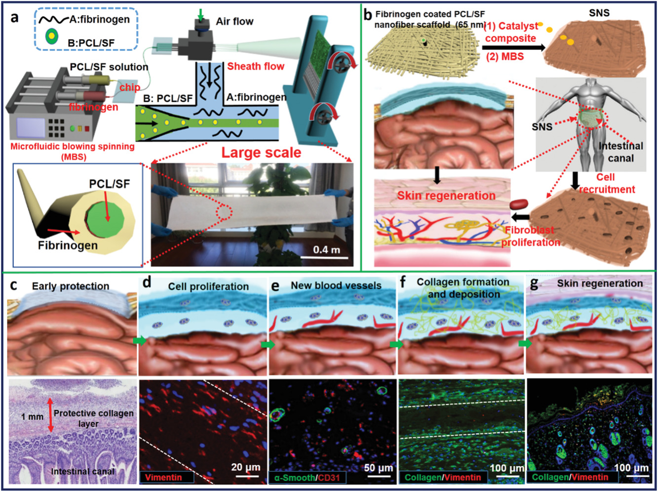
  • (a)准备生物可降解的纤维蛋白原覆盖的PCL/SF纳米纤维支架的示意图,通过MBS生成核壳结构的纳米纤维。
  • (b)腹部皮肤再生的3个阶段:可降解的SNS制造,皮肤组织重建和皮肤形成。其中,纤维蛋白原由MBS吹催化剂复合物(包含凝血酶),形成纤维蛋白。
  • (c-g)上图为腹壁缺损皮肤重建的示意图,下图为相关试验结果。
    • (c)早期形成胶原蛋白层的保护机制。
    • (d)细胞增殖。其中,波形蛋白(vimentin)是成纤维细胞的特异性蛋白。
    • (e)新生血管。其中,血小板-内皮细胞粘附分子(PECAM/CD31)α-平滑肌肌动蛋白(α-Smooth)用于定位血管。
    • (f)胶原蛋白的形成与沉积。
    • (g)皮肤更新。

  白色线框区域代表SNS区域。

Figure 2. Characterizations of nanofiber scaffolds and formation of SNS

  • (a-c)不同纺丝技术的扫描电镜图和纳米纤维直径分布。
    • (a)微流控吹塑。扫描电镜显示其具备多孔结构。
    • (b)静电纺丝。
    • (c)微流控纺丝。
  • (d)比较四种纺丝方法(静电纺丝、溶液吹塑、微流控纺丝、微流控吹塑)的纳米纤维直径。
  • (e)纤维蛋白原覆盖的纳米纤维支架的水接触角时间演变曲线(上)和图像(下)。
  • (f)纤维蛋白原覆盖的PCL/SF纳米纤维支架(左)和SNS(右)的数码照片。
  • (g)纳米纤维支架的扫描电镜图片。
  • (h)SNS的纳米纤维聚集在一起(由于胶体融合)的SEM图像(左,中)和SNS的荧光双染色图像(右)。
  • (i)PCL/SF纳米纤维表面凝血酶与纤维蛋白原形成SNS的反应机制及成纤维细胞的迁移。纤维蛋白原在催化剂复合物(Ca2+和凝血酶)的作用下,凝血因子VII激活,生成纤维蛋白单体,随后凝胶化形成纤维蛋白网络。

水接触角的作用:衡量润湿程度,即判断材料是否亲水。一般小于90°认为亲水。

Figure 3. In vivo study of the process (fibroblast proliferation and migration, normal connective tissue formation, collagen deposition, and neovascularization) of skin tissue reconstruction

  • (a)后背有大面积伤口的小鼠照片。
  • (b)经过SNS处理的伤口表面的流血的小血管止血的示意图。
  • (c)SNS植入大鼠背部受损区域1、3、5和7天后(红色箭头表示血管)。
  • (d)大鼠肝出血模型:十字切割大鼠肝(左),肝出血(右上),SNS止血(右下)。
  • (e)SNS植入1、3、5、7天的HE染色。红线指示SNS区域,紫色、黑色和蓝色箭头分别指示成纤维细胞,成纤维细胞迁移至SNS区域和胶原沉积。
  • (f)SNS植入1、3、5、7天的Masson染色。
  • (g)SNS植入1、3、5、7天的荧光染色。

Masson染色是指用两种或三种阴离子染料混合,胶原纤维呈蓝色,肌纤维呈红色。用来显示组织中纤维以及炎性因子的染色方法之一。

Figure 4. H&E staining (1, 3, 5), inflammatory cell fluorescent staining (2), vessels fluorescent staining (4), and collagen fluorescent staining (6) of the tissues around the implantation sites during different periods

  • (a)PCL/SF纳米纤维支架系统。红色箭头指炎症细胞,蓝色箭头指疤痕组织,绿色箭头指新生血管。纤维刺激巨噬细胞和中性粒细胞,显示可能会导致过度的机体免疫。此外,不能促进血管在膜对面再生。
  • (b)纤维蛋白密封胶组获得了均匀的血管分布。
  • (c)FS@PCL/SF组在血管分布上和PCL/SF组相似。暴露的纤维刺激了组织,导致了轻微地组织增生。
  • (d)SNS组中血管在SNS两边形成。只有本组胶原蛋白排列规律,分布均匀。白框代表正常组织。

Figure 5. Three processes (wound healing area, granulation growth thickness, and hair follicle formation) of skin formation
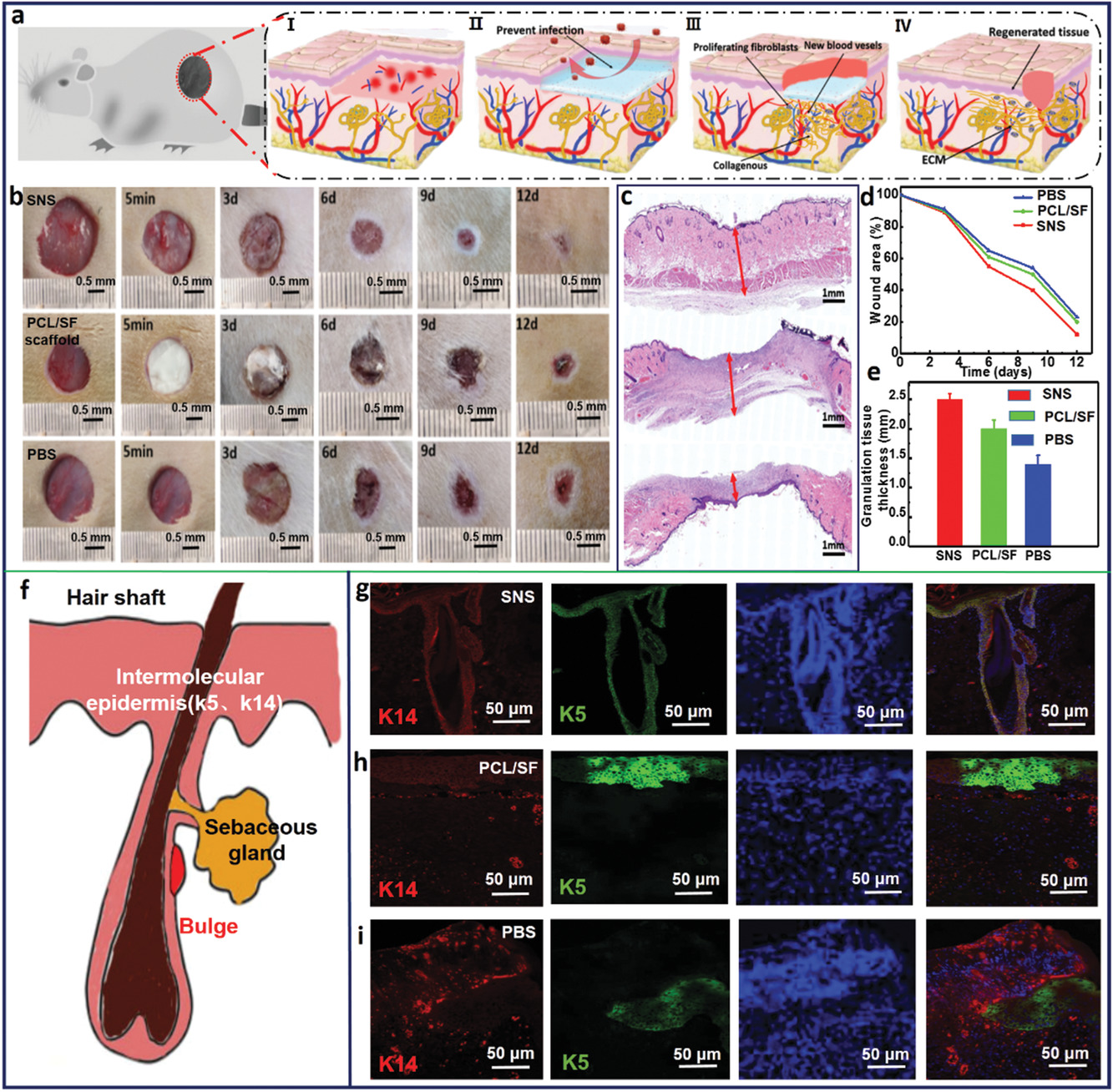
  • (a)伤口愈合过程的示意图。
  • (b)经SNS、PCL/SF纳米纤维支架和PBS处理的大鼠的代表性的皮肤伤口愈合过程的数码照片。
  • (c)SNS、PCL/SF纳米纤维支架和PBS组的肉芽生长的HE染色图。
  • (d)伤口区域面积和时间曲线图。
  • (e)肉芽组织厚度的定量分析。
  • (f)毛囊结构的草图。
  • (g-i)SNS、PCL/SF纳米纤维支架和PBS组的新生组织的K14和K5的双重荧光染色。
    • (g)可以看出有完整的新生组织毛囊结构。
    • (h)只形成了上皮而没有毛囊组织。
    • (i)新生组织结构模糊。

Figure 6. Application of SNS

  • (a)早期保护和组织再生的示意图。
  • (b)SNS和聚丙烯(PP)网组的新生组织和暴露的肠管的Mason染色。红色边界代表受伤区域,黑色边界代表SNS或PP网覆盖的区域。
  • (c)SNS组(上)和PP网组(下)暴露的肠管。红色边界代表SNS或PP网覆盖的区域。
  • (d-e)SNS和PP网处理的腹壁缺损的HE染色和天狼猩红染色:新血管(红色箭头标记)(左);新生组织的新组织结构(中)和胶原纤维(右)。
    • (d)SNS组的真皮里有大量微血管;有平滑的表面,即浆膜层和平滑肌层;有丰富的均匀的Ⅰ、Ⅱ胶原排列。
    • (e)PP网组观察不到微血管,平滑肌破裂,浆膜层破坏和胶原纤维紊乱,表明肠壁可能受损。
  • (f-g)分别植入人造皮肤和PP网的腹壁缺损的再生组织中的IL-6和肿瘤坏死因子TNF-α的免疫荧光染色。
    • (f)较少的IL-6和TNF-α被检测,显示轻微的炎性反应。
    • (g)显示严重的炎性反应。

天狼猩红(Picrosirius Red )染色可用于区分Ⅰ、Ⅱ、Ⅲ和Ⅳ型胶原,由于天狼猩红是一种强酸性阴离子染料,可与呈碱性的胶原反应,使胶原纤维在偏振光照射时,产生明显的双折光现象。不同的胶原纤维具有不同的旋光性,在偏振光显微镜下呈现相异的颜色,借以区分Ⅰ、Ⅱ、Ⅲ和Ⅳ型胶原,Ⅰ型纤维:紧密排列, 显示很强的双折光性,呈黄色或红色的纤维;Ⅱ型纤维:显示弱的双折光,呈多种色彩的疏松网状分布;Ⅲ型纤维:显示弱的双折光,呈绿色的细纤维;Ⅳ型纤维:显示弱的双折光的基膜,呈淡黄色。

天狼猩红染色可直接观察分析胶原纤维化程度,主要应用于各种组织病变时对胶原纤维异常或纤维增生的研究中。

Reference

Cui T, Yu J, Li Q, et al. Large-Scale Fabrication of Robust Artificial Skins from a Biodegradable Sealant-Loaded Nanofiber Scaffold to Skin Tissue via Microfluidic Blow-Spinning[J]. Advanced Materials, 2020, 32(32): e2000982.

评论

3D cell culture 3D cell culturing 3D cell microarrays 3D culture 3D culture model 3D printing 3D spheroid 3D tumor culture 3D tumors 3D vascular mapping ACT ADV AUTODESK Abdominal wall defects Acoustofluidics Adipocyte Adipogenesis Adoptive cell therapy AirPods Alginate Anticancer Anticancer agents Anticancer drugs Apple Apriori Association Analysis August AutoCAD Autodock Vina Bio-inspired systems Biochannels Bioengineering Bioinspired Biological physics Biomarkers Biomaterial Biomaterials Biomimetic materials Biomimetics Bioprinting Blood purification Blood-brain barrier Bone regeneration Breast cancer Breast cancer cells Breast neoplasms CM1860 CRISPR/Cas9 system CSS CTC isolation CTCs Cancer Cancer angiogenesis Cancer cell invasion Cancer immunity Cancer immunotherapy Cancer metabolism Cancer metastasis Cancer models Cancer screening Cancer stem cells Cell adhesion Cell arrays Cell assembly Cell clusters Cell culture Cell culture techniques Cell mechanical stimulation Cell morphology Cell trapping Cell-bead pairing Cell-cell interaction Cell-laden gelatin methacrylate Cellular uptake Cell−cell interaction Cervical cancer Cheminformatics Chemotherapy Chimeric antigen receptor-T cells Chip interface Circulating tumor cells Clinical diagnostics Cmder Co-culture Coculture Colon Colorectal cancer Combinatorial drug screening Combinatorial drug testing Compartmentalized devices Confined migration Continuous flow Convolutional neural network Cooking Crawler Cryostat Curved geometry Cytokine detection Cytometry Cytotoxicity Cytotoxicity assay DESeq DNA tensioners Data Mining Deep learning Deformability Delaunay triangulation Detective story Diabetic wound healing Diagnostics Dielectrophoresis Differentiation Digital microfluidics Direct reprogramming Discrimination of heterogenic CTCs Django Double emulsion microfluidics Droplet Droplet microfluidics Droplets generation Droplet‐based microfluidics Drug combination Drug efficacy evaluation Drug evaluation Drug metabolism Drug resistance Drug resistance screening Drug screening Drug testing Dual isolation and profiling Dynamic culture Earphone Efficiency Efficiency of encapsulation Elastomers Embedded 3D bioprinting Encapsulation Endothelial cell Endothelial cells English Environmental hazard assessment Epithelial–mesenchymal transition Euclidean distance Exosome biogenesis Exosomes Experiment Extracellular vesicles FC40 FP-growth Fabrication Fast prototyping Fibroblasts Fibrous strands Fiddler Flask Flow rates Fluorescence‐activated cell sorting Functional drug testing GEO Galgame Game Gene Expression Profiling Gene delivery Gene expression profiling Gene targetic Genetic association Gene‐editing Gigabyte Glypican-1 GoldenDict Google Translate Gradient generator Growth factor G‐CSF HBEXO-Chip HTML Hanging drop Head and neck cancer Hectorite nanoclay Hepatic models Hepatocytes Heterotypic tumor HiPSCs High throughput analyses High-throughput High-throughput drug screening High-throughput screening assays High‐throughput methods Histopathology Human neural stem cells Human skin equivalent Hydrogel Hydrogel hypoxia Hydrogels ImageJ Immune checkpoint blockade Immune-cell infiltration Immunoassay Immunological surveillance Immunotherapy In vitro tests In vivo mimicking Induced hepatocytes Innervation Insulin resistance Insulin signaling Interferon‐gamma Intestinal stem cells Intracellular delivery Intratumoral heterogeneity JRPG Jaccard coefficient JavaScript July June KNN Kidney-on-a-chip Lab-on-a-chip Laptop Large scale Lattice resoning Leica Leukapheresis Link Lipid metabolism Liquid biopsy Literature Liver Liver microenvironment Liver spheroid Luminal mechanics Lung cells MOE Machine Learning Machine learning Macro Macromolecule delivery Macroporous microgel scaffolds Magnetic field Magnetic sorting Malignant potential Mammary tumor organoids Manhattan distance Manual Materials science May Mechanical forces Melanoma Mesenchymal stem cells Mesoporous silica particles (MSNs) Metastasis Microassembly Microcapsule Microcontact printing Microdroplets Microenvironment Microfluidic array Microfluidic chips Microfluidic device Microfluidic droplet Microfluidic organ-on-a chip Microfluidic organ-on-a-chip Microfluidic patterning Microfluidic screening Microfluidic tumor models Microfluidic-blow-spinning Microfluidics Microneedles Micropatterning Microtexture Microvascular Microvascular networks Microvasculatures Microwells Mini-guts Mirco-droplets Molecular docking Molecular imprinting Monolith Monthly Multi-Size 3D tumors Multi-organoid-on-chip Multicellular spheroids Multicellular systems Multicellular tumor aggregates Multi‐step cascade reactions Myeloid-derived suppressor cells NK cell NanoZoomer Nanomaterials Nanoparticle delivery Nanoparticle drug delivery Nanoparticles Nanowell Natural killer cells Neural progenitor cell Neuroblastoma Neuronal cell Neurons Nintendo Nissl body Node.js On-Chip orthogonal Analysis OpenBabel Organ-on-a-chip Organ-on-a-chip devices Organically modified ceramics Organoids Organ‐on‐a‐chip Osteochondral interface Oxygen control Oxygen gradients Oxygen microenvironments PDA-modified lung scaffolds PDMS PTX‐loaded liposomes Pain relief Pancreatic cancer Pancreatic ductal adenocarcinoma Pancreatic islet Pathology Patient-derived organoid Patient-derived tumor model Patterning Pearl powder Pearson coefficient Penetralium Perfusable Personalized medicine Photocytotoxicity Photodynamic therapy (PDT) Physiological geometry Pluronic F127 Pneumatic valve Poetry Polymer giant unilamellar vesicles Polystyrene PowerShell Precision medicine Preclinical models Premetastatic niche Primary cell transfection Printing Protein patterning Protein secretion Pubmed PyMOL Pybel Pytesseract Python Quasi-static hydrodynamic capture R RDKit RNAi nanomedicine RPG Reactive oxygen species Reagents preparation Resistance Review Rod-shaped microgels STRING Selective isolation Self-assembly Self-healing hydrogel September Signal transduction Silk-collagen biomaterial composite Similarity Single cell Single cells Single molecule Single-cell Single-cell RNA sequencing Single‐cell analysis Single‐cell printing Size exclusion Skin regeneration Soft lithography Softstar Spheroids Spheroids-on-chips Staining StarBase Stem cells Sub-Poisson distribution Supramolecular chemistry Surface chemistry Surface modification Switch T cell function TCGA Tanimoto coefficient The Millennium Destiny The Wind Road Thin gel Tissue engineering Transcriptome Transfection Transient receptor potential channel modulators Tropism Tubulogenesis Tumor environmental Tumor exosomes Tumor growth and invasion Tumor immunotherapy Tumor metastasis Tumor microenvironment Tumor response Tumor sizes Tumor spheroid Tumor-on-a-chip Tumorsphere Tumors‐on‐a‐chip Type 2 diabetes mellitus Ultrasensitive detection Unboxing Underlying mechanism Vascularization Vascularized model Vasculature Visual novel Wettability Windows Terminal Word Writing Wuxia Xenoblade Chronicles Xin dynasty XuanYuan Sword Youdao cnpm fsevents miR-125b-5p miR-214-3p miRNA signature miRanda npm
Your browser is out-of-date!

Update your browser to view this website correctly. Update my browser now

×