Microfluidic tumor-on-a-chip model to evaluate the role of tumor environmental stress on NK cell exhaustion

Microfluidic tumor-on-a-chip model to evaluate the role of tumor environmental stress on NK cell exhaustion

  一种微流控肿瘤芯片,以评估NK细胞如何响应肿瘤诱导的抑制性环境。

Introduction

  在过去数年,多种免疫疗法突破如嵌合抗原受体(CAR)T细胞/NK细胞和免疫检查点抑制剂(ICIs)已经证明在血液癌(如急性髓系白血病和急性淋巴细胞白血病)中取得巨大成功。然而在将其应用到实体瘤中进展缓慢,部分原因可能是实体瘤产生的免疫抑制微环境压力。在这个背景下,最近的研究显示了肿瘤相关环境因子如营养不足、缺氧、废物累积和pH酸化抑制T和NK细胞抗肿瘤活性。因此理解这种微环境压力对免疫细胞的后果对于发展更有效的免疫治疗是非常关键的。然而使用传统的2D模型达到实体瘤微环境的完整性仍具挑战性。

  作者的肿瘤芯片模型包括一个微腔,在其中乳腺癌细胞被嵌入一个3D基质。位于微腔一端的内腔排列着内皮细胞,灌注培养基以滋养细胞,模仿肿瘤中的脉管系统。他们的结果证明了NK细胞最初展示了相当的细胞毒性能力,消灭了相当大比例的肿瘤细胞。然而培养在装置中逐渐侵蚀了它们的细胞毒性能力,导致了NK细胞耗竭。此外,作者探索了NK细胞的可塑性和它们扭转免疫耗竭的能力。在这个背景下,结果证明了NK细胞不能从耗竭表型中扭转,显示了尽管移除了肿瘤产生的环境压力,它们发生了多分子和功能改变。

Fig. 1. Tumor-on-a-chip

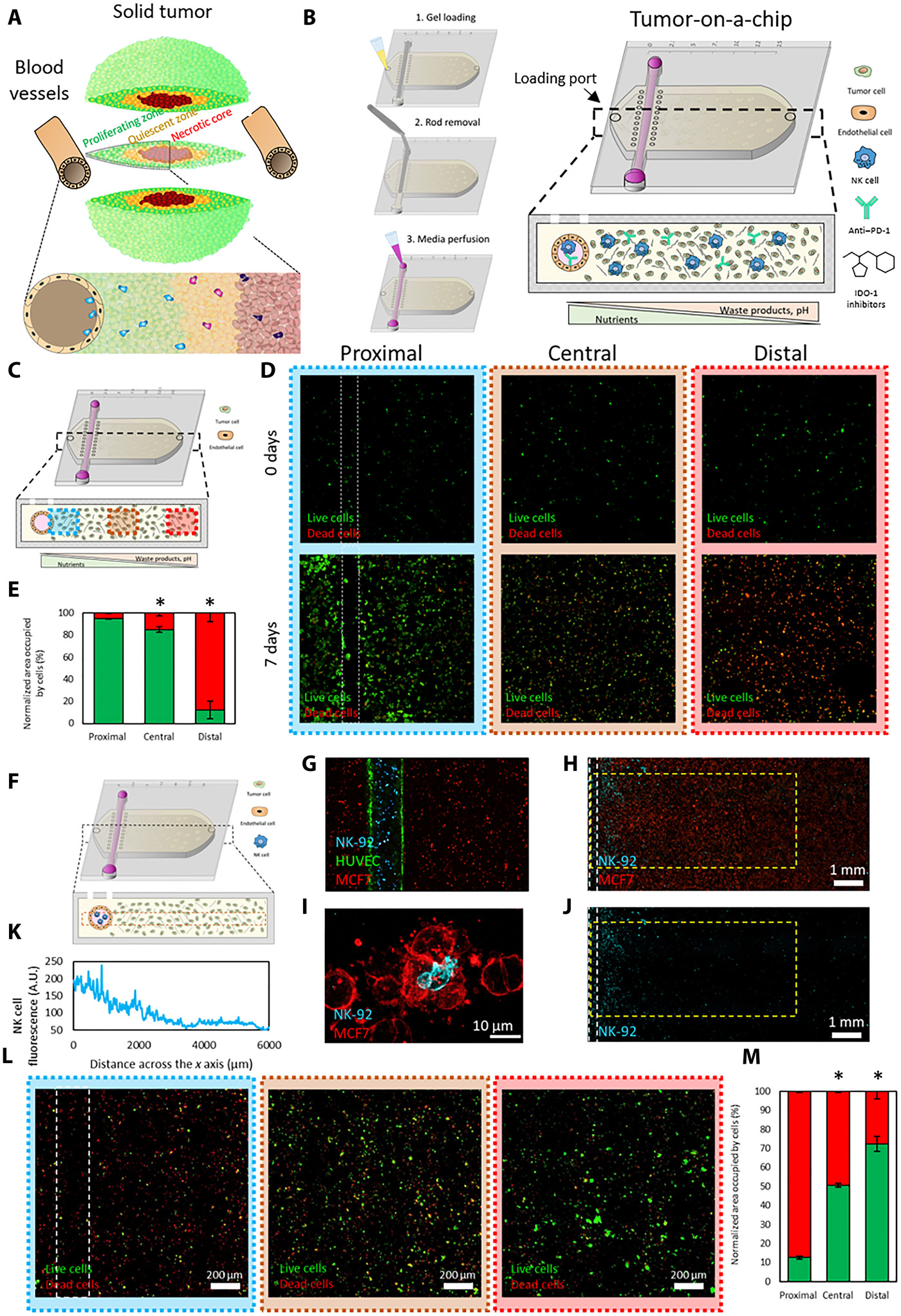
  • (A)由于营养不足导致了实体瘤中不同的肿瘤表型的示意图。
  • (B)肿瘤芯片的示意图。下方小图展示了微装置的截面。内腔排列着内皮细胞(如HUVECs)以代替血管,允许灌注培养基、NK-92细胞、PD-L1抗体(如阿特朱单抗)或IDO-1抑制剂(如epacadostat)。
  • (C)微装置的示意图, 近端、中央和远端区域分别显示为蓝色、橙色和红色。
  • (D)共聚焦显微图像显示了0天和7天的存活(绿)与死亡(红)的MCF7细胞。
  • (E)近端,中央和远端区域中被活细胞(绿色)和死细胞(红色)占据的区域。
  • (F)实验流程示意图。
  • (G)共聚焦显微图像显示了肿瘤芯片上的细胞扩散。MCF7细胞(红色)被嵌入胶原凝胶,NK-92细胞(蓝色)和HUVEC细胞(绿色)则嵌在内腔。
  • (H)共聚焦显微图像显示了NK-92细胞(蓝色)迁移到腔室和MCF7细胞(红色)。
  • (I)共聚焦图像展示了一个NK-92细胞与一个MCF7细胞交战。
  • (J)共聚焦图像突出了NK细胞迁移出内腔,进入腔室。
  • (K)通过测量NK细胞荧光定量NK-92细胞的x轴迁移。
  • (L)由于迁移效果一般,因此将MCF-7和NK-92细胞共培养一周。由于NK细胞接近营养丰富的内腔,近端区域有更高比例的死细胞。
  • (M)定量分析显示与内腔的距离和存活癌细胞(绿色)的数目是成比例的。

Fig. 2. Culture in the tumor-on-a-chip microdevice led to NK cell exhaustion

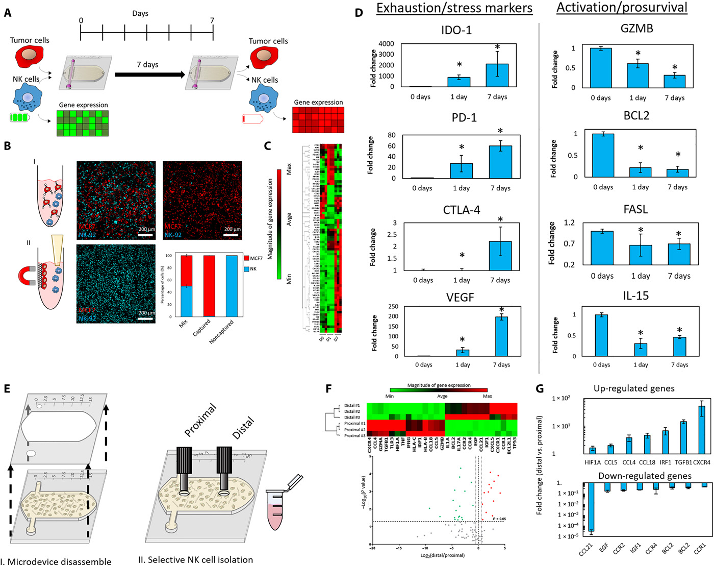
  • (A)实验测量免疫耗竭的示意图。NK细胞和MCF7细胞被混合入胶原,以1:3的比例(0.5 million cells / 1 ml:1.5 million cells / 1 ml)共培养7天。这个方法确保NK细胞在水凝胶中保持均匀密度。7天后,分离NK细胞,测量基因表达和其他特征。
  • (B)NK细胞分离示意图。MCF7连接到磁珠,由此可以分离NK细胞。共聚焦图像显示了捕获的MCF7细胞(红色)和分离的NK细胞(蓝色)。柱形图突出显示了NK细胞分离的效率。
  • (C)聚类图描绘了0天、1天和7天的基因表达。
  • (D)柱状图显示了耗竭标记物和激活/促生存基因的上/下调。
  • (E)空间控制NK细胞从近端和远端分离的示意图。
  • (F)聚类图和火山图定量近端和远端区域的基因表达。
  • (G)柱状图描绘了远端区域相比于近端区域增加的上调基因和减少的下调基因。

Fig. 3. Comparison of NK cell exhaustion in the tumor-on-a-chip microdevice with traditional 3D culture

  • (A)测量免疫耗竭实验的示意图。NK细胞和MCF7细胞以1:3的比例在孔板中共培养7天。7天后分离NK细胞,测量基因表达和其他特征。这个实验显示了在孔板而不是肿瘤芯片上的相似的耗竭参数。确定了NK细胞耗竭是由芯片里的环境压力引起的,而不是仅由肿瘤细胞存在7天的慢性激活引起的。
  • (B)聚类图显示了肿瘤芯片与孔板实验的基因表达变化。
  • (C-D)柱状图描绘了肿瘤芯片与孔板相比,耗竭标记物和激活/促生存基因的变化。
  • (E)蜘蛛图显示了肿瘤芯片相比于传统的孔板实验差异更大的10个基因通路。NRMOP:negative regulation of multicellular organismal process,多细胞生物过程的负调控;PRISP:positive regulation of immune system process,免疫系统过程的正调控;PRCA:positive regulation of cell activation,细胞激活的正调控;PRCP:positive regulation
    of cytokine production,细胞因子产生的正调控。这些通路被GSEA确定为为受影响最大的途径。

Fig. 4. Effect of ICIs and IDO-1 inhibitors in traditional assays and the tumor-on-a-chip

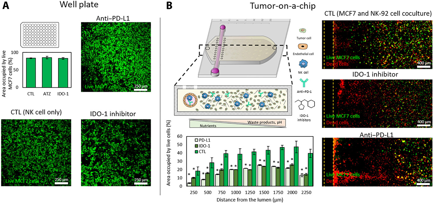
  • (A)在传统孔板上评估atezolizumab(PD-L1抗体)和epacadostat(IDO-1抑制剂)的潜在作用。汇合的单层MCF7被接种到96孔板,24小时后添加有/无IDO-1抑制剂或PD-L1抗体的NK-92细胞(9 MCF7:1 NK)。共聚焦图像显示NK-92细胞毒性潜能没有显著性改善。其中,NK细胞没有环境压力,并且没有显示相关通路的上调。
  • (B)相似的试验在肿瘤芯片上评估atezolizumab和epacadostat。共聚焦图像证明了IDO-1抑制剂或PD-L1抗体都增加了MCF7细胞坏死。死细胞(红色)集中在内腔近端,活的MCF7细胞(绿色)保持着远端区域。

Fig. 5. NK cell recovery from the tumor-on-a-chip platform

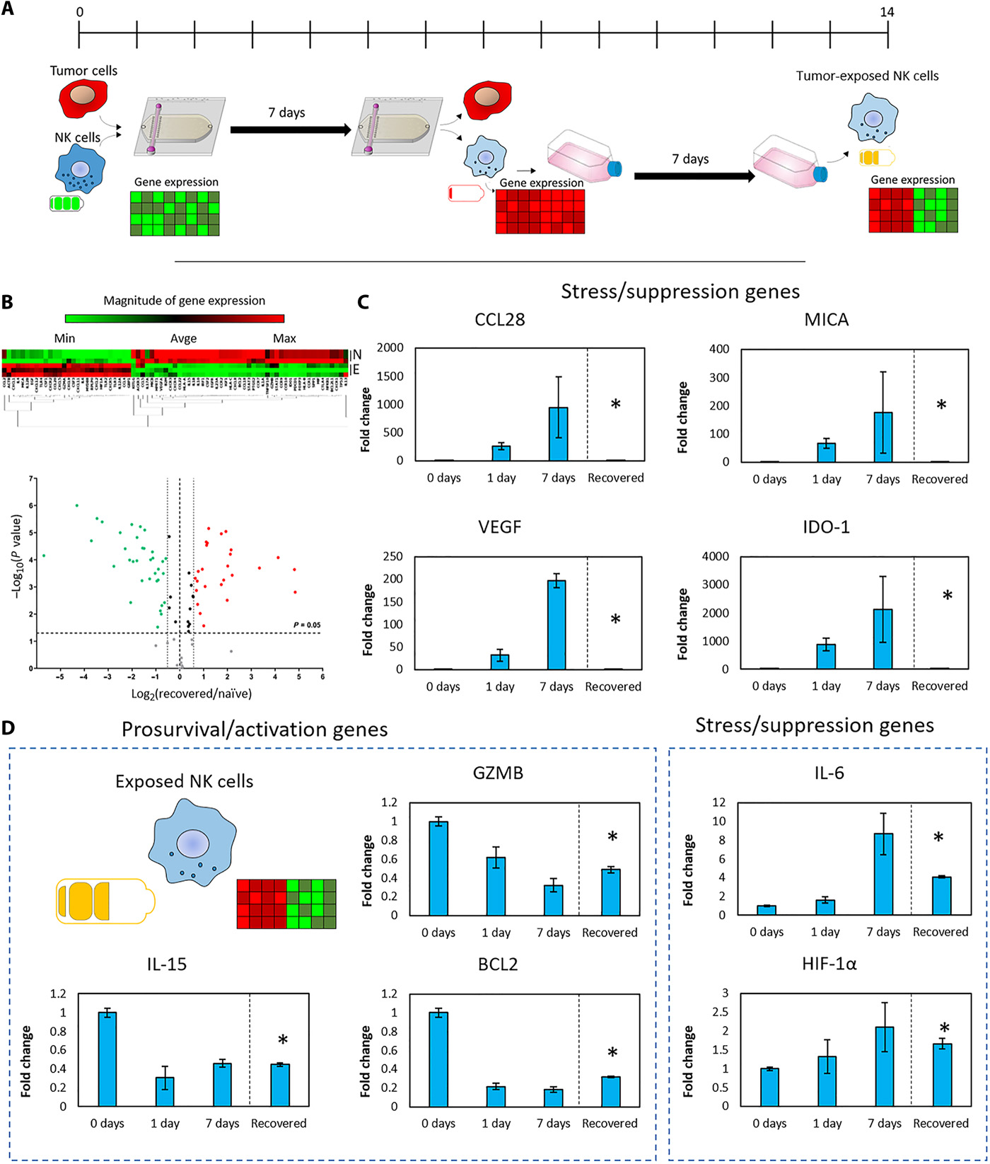
  • (A)评估NK-92细胞的恢复能力的实验示意图。NK-92细胞和MCF7细胞共培养(0.5 million NK:1.5 million MCF7 cells/ml)7天。随后,分离NK-92细胞,在传统培养瓶中额外培养一周并分析它们的基因表达。
  • (B)暴露NK细胞与天然NK细胞的基因表达聚类和火山图。
  • (C)柱状图显示多个在肿瘤芯片中上/下调的基因在1周后恢复到基准表达。
  • (D)柱状图显示与天然NK细胞相比,基因部分或没有恢复到基准表达。

Fig. 6. Functional changes in exposed NK cells

  • (A)从肿瘤芯片上分离的暴露NK细胞在1周的传统培养瓶培养后展示了多个基因表达的变化。
  • (B)光学代谢成像用于可视化细胞内NAD(P)H和FAD荧光强度[氧化还原比=NAD(P)H强度除以FAD强度]和平均荧光持续期(τm)。
  • (C)小提琴图显示了基于NAD(P)H和FAD强度的NK细胞氧化还原比。暴露的NK细胞与天然NK细胞相比有更少的氧化还原比,指示代谢重组。此外,暴露的NK细胞显示减少的NAD(P)H τm与增多的FAD τm。
  • (D)小提琴图显示氧化还原比、NAD(P)H τm和FAD τm是由不同代谢抑制剂与趋化因子导致:2DG,糖酵解抑制剂;依托莫昔(ETO),脂肪酸氧化抑制剂;寡霉素(Oligo),电子传递链复合体V抑制剂;DMSO,二甲基亚砜。
  • (E-F)延时显微镜分析暴露NK细胞在3D胶原水凝胶中迁移。独立的NK细胞轨迹描述为不同颜色。小提琴图显示暴露的NK细胞迁移速度更慢。
  • (G-H)暴露和天然NK细胞的细胞毒性分析。单层的MCF7接种到传统孔板,天然或暴露NK细胞添加到顶部。共聚焦图像显示二者的杀伤能力。
  • (I)柱状图分析显示不同比例的肿瘤细胞(TC)与NK细胞(NK)在(H)中的MCF7细胞的破坏。暴露的NK细胞显示更少的细胞毒性能力。

Fig. 7. NK cell dysfunction marker comparison

  • (A)体内功能失调(如耗竭和失活)的NK细胞的分子改变示意图。
  • (B)示意图描绘了模型中与MCF7细胞(接种密度:1.5 million cells/ml)共培养了7天的NK-92细胞(接种密度:0.5 million cells/ml)的变化。

Reference

Ayuso J M, Rehman S, Virumbrales-Munoz M, et al. Microfluidic tumor-on-a-chip model to evaluate the role of tumor environmental stress on NK cell exhaustion[J]. Science Advances, 2021, 7(8): eabc2331.

评论

3D cell culture 3D cell culturing 3D cell microarrays 3D culture 3D culture model 3D printing 3D spheroid 3D tumor culture 3D tumors 3D vascular mapping ACT ADV AUTODESK Abdominal wall defects Acoustofluidics Adipocyte Adipogenesis Adoptive cell therapy AirPods Alginate Anticancer Anticancer agents Anticancer drugs Apple Apriori Association Analysis August AutoCAD Autodock Vina Bio-inspired systems Biochannels Bioengineering Bioinspired Biological physics Biomarkers Biomaterial Biomaterials Biomimetic materials Biomimetics Bioprinting Blood purification Blood-brain barrier Bone regeneration Breast cancer Breast cancer cells Breast neoplasms CM1860 CRISPR/Cas9 system CSS CTC isolation CTCs Cancer Cancer angiogenesis Cancer cell invasion Cancer immunity Cancer immunotherapy Cancer metabolism Cancer metastasis Cancer models Cancer screening Cancer stem cells Cell adhesion Cell arrays Cell assembly Cell clusters Cell culture Cell culture techniques Cell mechanical stimulation Cell morphology Cell trapping Cell-bead pairing Cell-cell interaction Cell-laden gelatin methacrylate Cellular uptake Cell−cell interaction Cervical cancer Cheminformatics Chemotherapy Chimeric antigen receptor-T cells Chip interface Circulating tumor cells Clinical diagnostics Cmder Co-culture Coculture Colon Colorectal cancer Combinatorial drug screening Combinatorial drug testing Compartmentalized devices Confined migration Continuous flow Convolutional neural network Cooking Crawler Cryostat Curved geometry Cytokine detection Cytometry Cytotoxicity Cytotoxicity assay DESeq DNA tensioners Data Mining Deep learning Deformability Delaunay triangulation Detective story Diabetic wound healing Diagnostics Dielectrophoresis Differentiation Digital microfluidics Direct reprogramming Discrimination of heterogenic CTCs Django Double emulsion microfluidics Droplet Droplet microfluidics Droplets generation Droplet‐based microfluidics Drug combination Drug efficacy evaluation Drug evaluation Drug metabolism Drug resistance Drug resistance screening Drug screening Drug testing Dual isolation and profiling Dynamic culture Earphone Efficiency Efficiency of encapsulation Elastomers Embedded 3D bioprinting Encapsulation Endothelial cell Endothelial cells English Environmental hazard assessment Epithelial–mesenchymal transition Euclidean distance Exosome biogenesis Exosomes Experiment Extracellular vesicles FC40 FP-growth Fabrication Fast prototyping Fibroblasts Fibrous strands Fiddler Flask Flow rates Fluorescence‐activated cell sorting Functional drug testing GEO Galgame Game Gene Expression Profiling Gene delivery Gene expression profiling Gene targetic Genetic association Gene‐editing Gigabyte Glypican-1 GoldenDict Google Translate Gradient generator Growth factor G‐CSF HBEXO-Chip HTML Hanging drop Head and neck cancer Hectorite nanoclay Hepatic models Hepatocytes Heterotypic tumor HiPSCs High throughput analyses High-throughput High-throughput drug screening High-throughput screening assays High‐throughput methods Histopathology Human neural stem cells Human skin equivalent Hydrogel Hydrogel hypoxia Hydrogels ImageJ Immune checkpoint blockade Immune-cell infiltration Immunoassay Immunological surveillance Immunotherapy In vitro tests In vivo mimicking Induced hepatocytes Innervation Insulin resistance Insulin signaling Interferon‐gamma Intestinal stem cells Intracellular delivery Intratumoral heterogeneity JRPG Jaccard coefficient JavaScript July June KNN Kidney-on-a-chip Lab-on-a-chip Laptop Large scale Lattice resoning Leica Leukapheresis Link Lipid metabolism Liquid biopsy Literature Liver Liver microenvironment Liver spheroid Luminal mechanics Lung cells MOE Machine Learning Machine learning Macro Macromolecule delivery Macroporous microgel scaffolds Magnetic field Magnetic sorting Malignant potential Mammary tumor organoids Manhattan distance Manual Materials science May Mechanical forces Melanoma Mesenchymal stem cells Mesoporous silica particles (MSNs) Metastasis Microassembly Microcapsule Microcontact printing Microdroplets Microenvironment Microfluidic array Microfluidic chips Microfluidic device Microfluidic droplet Microfluidic organ-on-a chip Microfluidic organ-on-a-chip Microfluidic patterning Microfluidic screening Microfluidic tumor models Microfluidic-blow-spinning Microfluidics Microneedles Micropatterning Microtexture Microvascular Microvascular networks Microvasculatures Microwells Mini-guts Mirco-droplets Molecular docking Molecular imprinting Monolith Monthly Multi-Size 3D tumors Multi-organoid-on-chip Multicellular spheroids Multicellular systems Multicellular tumor aggregates Multi‐step cascade reactions Myeloid-derived suppressor cells NK cell NanoZoomer Nanomaterials Nanoparticle delivery Nanoparticle drug delivery Nanoparticles Nanowell Natural killer cells Neural progenitor cell Neuroblastoma Neuronal cell Neurons Nintendo Nissl body Node.js On-Chip orthogonal Analysis OpenBabel Organ-on-a-chip Organ-on-a-chip devices Organically modified ceramics Organoids Organ‐on‐a‐chip Osteochondral interface Oxygen control Oxygen gradients Oxygen microenvironments PDA-modified lung scaffolds PDMS PTX‐loaded liposomes Pain relief Pancreatic cancer Pancreatic ductal adenocarcinoma Pancreatic islet Pathology Patient-derived organoid Patient-derived tumor model Patterning Pearl powder Pearson coefficient Penetralium Perfusable Personalized medicine Photocytotoxicity Photodynamic therapy (PDT) Physiological geometry Pluronic F127 Pneumatic valve Poetry Polymer giant unilamellar vesicles Polystyrene PowerShell Precision medicine Preclinical models Premetastatic niche Primary cell transfection Printing Protein patterning Protein secretion Pubmed PyMOL Pybel Pytesseract Python Quasi-static hydrodynamic capture R RDKit RNAi nanomedicine RPG Reactive oxygen species Reagents preparation Resistance Review Rod-shaped microgels STRING Selective isolation Self-assembly Self-healing hydrogel September Signal transduction Silk-collagen biomaterial composite Similarity Single cell Single cells Single molecule Single-cell Single-cell RNA sequencing Single‐cell analysis Single‐cell printing Size exclusion Skin regeneration Soft lithography Softstar Spheroids Spheroids-on-chips Staining StarBase Stem cells Sub-Poisson distribution Supramolecular chemistry Surface chemistry Surface modification Switch T cell function TCGA Tanimoto coefficient The Millennium Destiny The Wind Road Thin gel Tissue engineering Transcriptome Transfection Transient receptor potential channel modulators Tropism Tubulogenesis Tumor environmental Tumor exosomes Tumor growth and invasion Tumor immunotherapy Tumor metastasis Tumor microenvironment Tumor response Tumor sizes Tumor spheroid Tumor-on-a-chip Tumorsphere Tumors‐on‐a‐chip Type 2 diabetes mellitus Ultrasensitive detection Unboxing Underlying mechanism Vascularization Vascularized model Vasculature Visual novel Wettability Windows Terminal Word Writing Wuxia Xenoblade Chronicles Xin dynasty XuanYuan Sword Youdao cnpm fsevents miR-125b-5p miR-214-3p miRNA signature miRanda npm
Your browser is out-of-date!

Update your browser to view this website correctly. Update my browser now

×